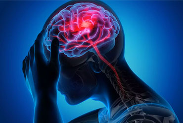
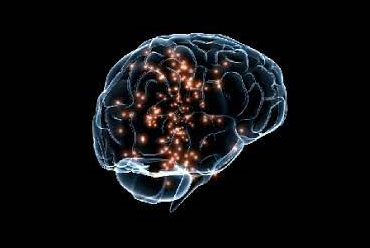
Expansion of the field of neurological treatment.
Along with the standardization of clinical neurology departments and the establishment of the specialist neurologist certification system, the treatment and research conducted by neurologists has increased dramatically, while, at the same time, new treatment methods for numerous neurological disorders that were previously considered refractory or untreatable have been developed. However, while these new treatment methods are being continuously developed, many aspects of the medical insurance system in Japan are not entirely appropriate for the dissemination and promotion of these new methods throughout society. This has been proactively engaged in activities to socially resolve the various problems associated with neurology treatment.
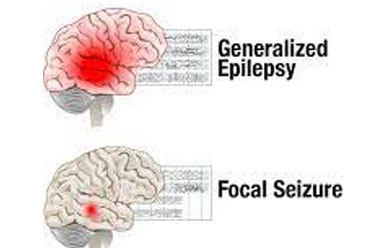
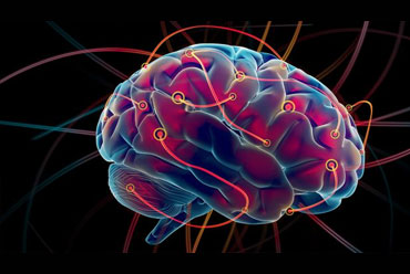
With diversification in the treatment of neurological diseases, increasing social demand for the creation of guidelines on the basis of evidence-based medicine has led to the establishment of an ad hoc committee for the creation of treatment guidelines for the six major diseases as Parkinson's disease, chronic headache, epilepsy, amyotrophic lateral sclerosis, dementia disorders, and stroke. As a result, treatment guidelines for the six major diseases were published over the period from May 2012 to May 2013. Since then, these guidelines have been revised or are currently under revision.
The sleep lab
Neurosciences Research Centre accommodates the Sleep Lab, the place where the study insomnia. “This is one of very few places where insomnia is researched so extensively and in such depth, by means of combining various cutting-edge methods.
This is one of very few places where insomnia is researched so extensively and in such depth, by means of combining various cutting-edge methods.”
Is it unusual to study insomnia? Yes and no. No, because poor sleepers are a worldwide subject of clinical study. “But those studies tend to focus on sleepers who are thought to suffer from a sleeping disorder, such as sleep apnea or narcolepsy,”. Yes, because the Sleep Lab is the only lab that carries out research into the most common sleeping disorder: “ordinary” poor sleep.
Some 10 percent of the population suffer from poor sleep. Insomnia as such is not deemed an official disease, the handbook for the classification of psychiatric disorders. The poor sleep may have very negative effects on an individual’s health, such as tiredness, exhaustion, depression, stress, cardiovascular disorders and obesity.
Central theme in this lab is the understanding of ocular signal transduction as it relies on the optical parts of the eye and retina. Optical signal transduction is characterized by the retinal point-spread-function. The transparency of the relevant biological components is a critical factor. How transparency is achieved and how defects can be characterized are important research questions of this lab. Normal as well as pathological conditions, including surgical and transplantation effects are investigated. We have specialized at the retina led to an instrument for practical clinical assessment of straylight glare sensitivity called C-Quant.
The study of eye movements not only addresses debilitating neuro- ophthalmological problems but has become an essential tool of basic neuroscience research. Eye movements have assumed another key role in neuroscience, as quantitative markers of higher-level aspects of behavior. Many studies that focus on cognitive and behavioral neuroscience—for example, reward, attention, planning, and prediction—and disorders, such as autism, schizophrenia, and neglect, use eye movements as a “read out” of what the brain is planning, mulling over, or excited about. Eye movements become an ideal tool to probe both function and malfunction of the cerebellum. Neurologists become more aware of how eye movements can be used in their clinics, for diagnosis, monitoring of therapies, and clinical and basic research. A careful clinical bedside evaluation, using only visual inspection, of each of the major subtypes of eye movements—saccades, pursuit, gaze holding, vestibular responses, and alignment and vergence—takes only a few minutes and provides much useful information toward accurate diagnosis.
Normal visual perception requires the proper functioning of ocular motor
systems that control the position and movement of the eyes to focus the image
of the object-of-interest (i.e., the visual target) on corresponding areas of
the retinas of the two eyes. For example, in addition to producing adjustments
in pupil size and lens refraction, accommodation involves the convergence of
the two eyes to direct onto the foveae the images of near objects. Eye
movements are also controlled to direct the eyes towards a visual target and to
follow the movements of the visual target. Such eye movements are controlled
by gaze systems. They coordinate the movement of the two eyes to ensure
that the images on the two retinas fall on corresponding areas of the binocular
field. When this fails, diplopia (double vision) results.
The extraocular muscles execute eye movements and are innervated by three cranial nerves. The muscles are attached to the sclera of the eye at one end and are anchored to the bony orbit of the eye at their opposite ends. Contraction of the muscles produce movement of the eyes within the orbit. The cranial lower motor neurons innervate these muscles and thereby control their contractions. For each eye, six muscles work together to control eye position and movement. Two extraocular muscles, the medial rectus and lateral rectus, work together to control horizontal eye movements.
The motor neurons controlling synergist and antagonist muscles must
coordinate their activities to produce the desired eye movements. Within the
abducens nucleus are abducens interneurons, which send their axons into
the contralateral medial longitudinal fasciculus. The abducens
interneurons coordinate the activity of the ipsilateral lateral rectus with that
of the contralateral medial rectus. Excitation of the interneurons in the left
abducens nucleus will excite neurons in the right oculomotor nucleus that
innervate the right medial rectus. Contraction of the right medial results in adduction of the right eye. Consequently, both the right and left eyes will be
directed towards the left when the left abducens nucleus is excited.
Interconnections between the trochlear nucleus and oculomotor nuclear complex coordinate their activity to allow the upward and downward movement of the eyes. These interconnecting axons appear to travel along with the fibers of the tectospinal tract
There are four basic types of eye movements: saccades, smooth pursuit movements, vergence movements , and vestibulo-ocular movements. Saccades are rapid, ballistic movements of the eyes that abruptly change the point of fixation. They range in amplitude from the small movements made while reading, for example, to the much larger movements made while gazing around a room. Saccades can be elicited voluntarily, but occur reflexively whenever the eyes are open, even when fixated on a target. Smooth pursuit movements are much slower tracking movements of the eyes designed to keep a moving stimulus on the fovea. Such movements are under voluntary control in the sense that the observer can choose whether or not to track a moving stimulus. Vergence movements align the fovea of each eye with targets located at different distances from the observer. Vestibulo-ocular movements stabilize the eyes relative to the external world, thus compensating for head movements. These reflex responses prevent visual images from “slipping” on the surface of the retina as head position varies. The action of vestibulo-ocular movements can be appreciated by fixating an object and moving the head from side to side; the eyes automatically compensate for the head movement by moving the same distance but in the opposite direction, thus keeping the image of the object at more or less the same place on the retina.